Foot & Heel Pain: Clinician-Led Self-Tests & Swedish Care Pathways
Persistent pain under the heel, behind the ankle, or across the ball of the foot frequently causes months of functional impairment. For expats and private patients in Sweden, understanding whether symptoms stem from plantar fasciitis, Achilles tendinopathy, or Morton’s neuroma is vital to avoid prolonged trial-and-error.
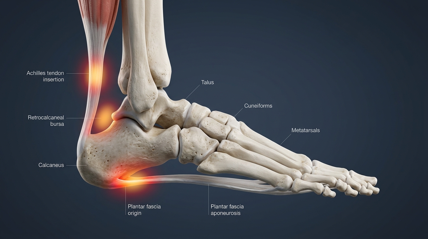
Where Does It Hurt? Three Distinct Pain Signatures
- Under the Heel (Plantar Surface): Characteristic of plantar fasciitis. Severe sharp pain during the first few steps in the morning or after prolonged sitting, often easing slightly after gentle movement before aching again in the evening.
- Posterior Heel & Ankle (Achilles Zone): Typical of Achilles tendinopathy or retrocalcaneal bursitis. Marked stiffness when climbing stairs, hill walking, or starting a running stride.
- Forefoot & Metatarsal Heads: Strongly suggestive of Morton’s neuroma or metatarsalgia. Sharp, shooting or burning sensations radiating into the third and fourth toes, often described as walking on a fold in your sock or a pebble.
Three Guided Orthopedic Self-Tests
Perform these movements gently on a level surface. Discontinue immediately if you experience acute shooting pain.
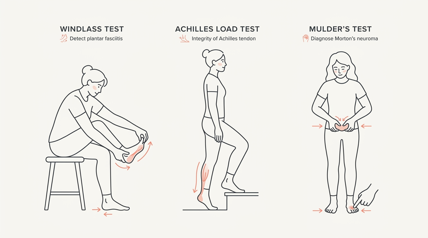
Test 1: Windlass Test (Plantar Fasciitis / Heel Spur)
Procedure: Sit with your affected leg crossed over your opposite knee. Grasp your big toe and pull it firmly upwards/backwards towards your shin while using your opposite thumb to press firmly on the inner underside of your heel bone (the medial calcaneal tubercle).
Positive Finding: Reproduction of sharp, localized tenderness under the heel as the plantar fascia tenses under load confirms mechanical strain consistent with plantar fasciitis.
Test 2: Single-Leg Heel Raise & Palpation (Achilles Tendinopathy)
Procedure: Stand on one leg on a step edge with your heel hanging off the edge. Perform a slow, controlled single-leg calf raise, and lower down below the step level. Next, gently pinch and palpate the Achilles tendon 2 to 6 cm above the heel bone.
Positive Finding: Pain during the eccentric lowering phase combined with localized tenderness and palpable thickening on the tendon body indicates mid-portion Achilles tendinopathy.
Test 3: Mulder’s Squeeze Test (Morton’s Neuroma)
Procedure: While seated, grasp across your forefoot with one hand and firmly squeeze the metatarsal bones together from side to side. Concurrently, press your thumb firmly from beneath between your third and fourth toes.
Positive Finding: A sudden sharp, electric shock radiating to the toes or a palpable click (Mulder’s click) strongly indicates intermetatarsal neuroma compression.
Differential Diagnosis Matrix
| Condition | Pain Pattern | Primary Aggravating Factor | Diagnostic Standard |
|---|---|---|---|
| Plantar Fasciitis | Stabbing morning heel pain; worsens after resting periods. | Barefoot walking on hardwood floors, first morning steps. | Clinical assessment + Ultrasound / MRI if unresolved after 6–8 weeks. |
| Achilles Tendinopathy | Morning stiffness and dull ache behind heel; warm-up phenomenon. | Uphill running, jumping, sudden eccentric loads. | Clinical load test + High-resolution Doppler ultrasound. |
| Morton’s Neuroma | Burning or electric shock sensation in the ball of the foot and toes. | Narrow shoes, high heels, repetitive forefoot impact. | Mulder's test + Dedicated Foot MRI or focused ultrasound. |
| Calcaneal / Metatarsal Stress Fracture | Progressive, localized deep bone ache that persists at rest; night pain. | All weight-bearing impact; does not ease with warm-up. | Magnetic Resonance Imaging (MRI) is required (plain X-rays miss early fractures). |
⚠️ Red Flags – When to Seek Emergency Care
If you heard or felt an audible "pop" or snapping sensation in the back of your ankle followed by inability to perform a single-leg heel raise (suspected complete Achilles tendon rupture), or if the foot exhibits sudden hot erythema, fever, or sensory loss with diabetes, contact emergency care or call 1177 immediately.
When Should You Consider a Private MRI in Sweden?
In the Swedish public system, accessing specialized MRI scans for foot and heel conditions frequently entails extensive primary care delays. If your pain has failed to resolve within 6 weeks of rehabilitation or if deep bone pathology is suspected, a private Foot & Ankle MRI provides decisive clarity:
- Detects occult bone stress injuries (bone marrow edema) weeks before visible on plain radiograph.
- Differentiates partial thickness plantar fascia tears from chronic recalcitrant fasciosis.
- Delivers exact millimeter measurement of neuromas for targeted intervention.
Obtain an Expedited Clinical Diagnosis & Rehabilitation Protocol
Schedule a tele-physiotherapy evaluation within 24 hours (SEK 690), or secure our comprehensive private orthopedic investigation bundle with guaranteed MRI within 10 days (SEK 9,900).
Book at Vård.se →